



Disclaimer: Early release articles are not considered as final versions. Any changes will be reflected in the online version in the month the article is officially released.
Author affiliation: Centre hospitalier d’Arras, Arras, France (J.-M. Devaux, J. Fleuriet, C. Leroy, K. Delaforge); Université de Rennes, INSERM, EHESP, IRSET, and Centre hospitalier universitaire de Rennes, Rennes, France (J.-P. Gangneux, B. Autier)
In 2026, a woman in her late 20s, originally from Kinshasa, Democratic Republic of the Congo, sought care for abdominal pain without fever in the emergency department of Arras Hospital Center in northern France. A physical examination showed localized abdominal erythema, and the emergency physician suspected bacterial cellulitis. We initiated oral amoxicillin/clavulanic acid (1 g 3×/d) for 7 days. Two weeks after her initial visit to the emergency department, she returned with persistent pain and a periumbilical mass, raising suspicion of a strangulated umbilical hernia. She was admitted for surgical management.
Figure
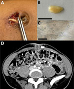
Figure. Armillifer armillatusparasite imaging in case of human infection mimicking strangulated umbilical hernia, France, 2026. A) Perioperative macroscopic photograph of the umbilical subcutaneous cyst and parasite. B) Gross appearance of…
Surgical exploration did not reveal a hernia but did reveal a white worm-like specimen enclosed within a cyst (Figure, panel A). We sent the specimen to the laboratory for analysis. The cylindrical and annulated morphology together with its 2-cm length indicated the specimen was a pentastomid parasite (Figure, panel B), which was consistent with the patient’s country of origin. In addition, microscopic examination of the parasite showed a typical pentastomid body wall with cuticular openings (1) (Figure, panel C). We identified the species by partially sequencing 18S rRNA (367 bp) and cox-1 (224 bp) genes, as previously described (2). Both partial sequences showed 100% identity with Armillifer armillatus sequences from GenBank (accession nos. PZ452549 [18S rRNA] and PZ452654 [cox-1]). A thoracoabdominal computed tomography scan showed a 15-mm subcutaneous cyst with surrounding fat stranding (Figure, panel D). We detected no other lesions, including no C-shaped calcifications.
Further epidemiologic investigation revealed that the patient consumed snake meat in Kinshasa and frequently had snakes in her vegetable garden. The patient had moved from the Democratic Republic of the Congo to France when she was 15 years of age and reported no subsequent travel to A. armillatus–endemic areas or known exposure to snakes or imported food products. Although her history raises the possibility of long-term parasite persistence, Armillifer nymphs are generally thought to survive for only a few years before degenerating and calcifying. Moreover, we could not exclude a more recent exposure because of potential recall bias or incomplete reporting of exposure history. Therefore, the duration of infection and viability of the nymph at the time of excision remains uncertain. We did not administer antiparasitic treatment because we surgically extracted the only detected parasite and because no drug therapy has demonstrated efficacy against pentastomiasis.
A. armillatus is a vermiform parasite belonging to Pentastomida, an atypical taxon related to the subphylum Crustacea (1). Human visceral pentastomiasis is mainly caused by species belonging to the genera Armillifer (A. armillatus, A. grandis, and A. moniliformis), Linguatula (L. serrata), and Porocephalus (P. crotali). Among those species, A. armillatus is predominantly reported in West Africa, and A. grandis is mostly reported in Central Africa, whereas other species have different geographic distributions (1). Adult Armillifer spp. and P. crotali parasites infect the respiratory tract of snakes (the definitive hosts) and lay eggs that are released into the environment through bronchial secretions or feces. Mammals are intermediate hosts and become infected with larvae and then nymphs after ingesting the parasite eggs. Nymphs encyst within the tissues of intermediate hosts and transform into adults after ingestion by a snake. Humans are incidental hosts and are typically infected through ingestion of raw or undercooked snake meat or ingestion of parasite egg–contaminated food or water.
In humans, larvae migrate and encyst mostly in the peritoneum, liver parenchyma, or abdominal lymph nodes. Because humans are not normally consumed by definitive hosts, the parasitic life cycle is not completed. Encysted nymphs can remain in tissues for several years before degenerating and eventually becoming calcified, which produces the characteristic C-shaped calcifications visible during imaging (2). Subcutaneous localization is less frequently reported and might manifest as a painful abdominal wall mass, potentially mimicking a surgical emergency. Similar manifestations involving inguinal hernias have previously been described (1,3). Most infections are asymptomatic. Symptomatic cases are uncommon and might be caused by inflammatory reactions to degenerating larvae. A diagnosis is usually made according to imaging or histopathology, but species identification is confirmed by using molecular methods (4).
In conclusion, this case illustrates an unusual clinical manifestation of A. armillatus infection that mimicked a surgical emergency in an A. armillatus–nonendemic setting. The long interval between exposure and symptom onset suggests a prolonged asymptomatic infection had occurred. The mechanisms underlying delayed clinical manifestations remain unclear. With increasing travel and human migration, clinicians in regions that are not endemic for A. armillatus infections should be aware of this parasitic infection, particularly in patients with relevant exposure history. Molecular tools are essential for A. armillatus identification but morphologically based diagnostic orientation and procedures are also required.
Dr. Devaux is a medical biologist resident in the Department of Laboratory Medicine at the Centre hospitalier d’Arras in Arras, France. His research interests include clinical microbiology and tropical diseases.