



Disclaimer: Early release articles are not considered as final versions. Any changes will be reflected in the online version in the month the article is officially released.

Author affiliation: Hospital of the University of Pennsylvania, Philadelphia, Pennsylvania, USA (D.C. Sung, R. Colbourn, N. Khan, J. DiRienzi, L. Randolph, F. Valdivieso, S. Mohan, H.I. Chen, S. Barrios, K. Degnan, A. Lustig, A.H. Tatarian, N.A. Aqui, L. Glaser, L. Litzky, M.P. Nasrallah); Centers for Disease Control and Prevention, Atlanta, Georgia, USA (G. Ficarra, B. Leitgeb, C. Gigante, L. Orciari, M. Niezgoda, P. Yager, R. Wallace, J. Bhatnagar, J.A. Villalba); Pennsylvania Department of Health, Harrisburg, Pennsylvania, USA (K. Mason, P. Kesavaramanujam, H. Tagaram, B. Schroeder); Chester County Health Department, West Chester, Pennsylvania, USA (A. Schafer, K. Raum)
Powassan virus (POWV) is a tickborne flavivirus that causes severe encephalitis. Cases are increasing annually; 60 cases and 9 deaths were confirmed in the United States during 2024 (1). Neuroinvasive disease occurs in 94% of confirmed cases and 98% of fatalities (1). We describe novel histopathologic findings of disseminated POWV infection with multiorgan involvement in a liver transplant recipient in Pennsylvania, USA.
Figure
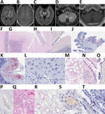
Figure. Magnetic resonance imaging and microphotographs for detection of POWV lineage 2 (deer tick virus) in postmortem tissues from a liver transplant patient with fatal infection and histopathological features of formalin-fixed,…
The patient was a 38-year-old woman in southeastern Pennsylvania, USA, who had alcohol-associated cirrhosis and remote preexposure prophylactic rabies vaccination 16 years earlier. She had undergone orthotopic liver transplant 6 weeks before seeking care at an emergency department for malaise, headache, fever, and leukocytosis. After admission, she had multiple generalized seizures. Magnetic resonance imaging revealed hydrocephalus with rapidly progressive enhancement of the cerebellum, brainstem, white matter, cortex, leptomeninges, and spinal cord (Figure, panels A–E). Cerebrospinal fluid (CSF) studies were consistent with viral encephalitis (Appendix Table 1). We ruled out autoimmune and other causes of viral encephalitis. Brain biopsy demonstrated lymphocytic encephalitis without features of a specific infectious etiology. We noted necrotic tissue intraoperatively, and the procedure was complicated by hemorrhage. The patient’s neurologic status deteriorated further, and she died after being transitioned to comfort care. CSF reverse transcription PCR testing for POWV was positive for lineage 2 (deer tick virus) the day before her death. POWV serum IgM test result was positive after her death (Table). Of note, CSF metagenomic sequencing on an earlier sample was negative for POWV (Appendix Table 1), which might reflect low viral load or testing detection limits.
Because of the patient’s rapid neurologic decline and imaging findings, rabies was initially suspected because other common causes were ruled out. Testing was expeditiously conducted for the safety of other organ recipients. Antemortem serum and CSF indirect immunofluorescent antibody rabies testing detected IgG, but not IgM. A rapid fluorescent focus inhibition test detected high serum rabies antibody titers and low CSF titers. Presence of rabies IgG in the CSF, although atypical, was attributed to previous vaccination and might have resulted from serum antibody spillover secondary to leaky blood–brain barrier from severe meningoencephalitis. Results of additional antemortem and postmortem rabies test results, including immunohistochemistry and PCR of the brain and liver, were negative (Appendix Table 2).
At autopsy, the brain contained a 3.2-cm focus of right basal ganglia necrosis and hemorrhage, diffuse edema, and bilateral basal ganglia softening. Histopathologic testing revealed widespread meningoencephalitis involving the cerebrum, pons, and cerebellum, with diffuse lymphoplasmacytic parenchymal and leptomeningeal inflammation, microgliosis, and marked Purkinje cell loss (Figure, panels F–H). Microglial nodules were not prominent, and intranuclear viral inclusions (Negri bodies) were absent. Available viral stain results were negative. POWV antigens and nucleic acids were detected by immunohistochemistry, in situ hybridization, or reverse transcription PCR (Table) in postmortem brain samples (Figure, panels I–L). Non–central nervous system findings included mild lymphocytic pneumonitis and bronchiolar epithelial apoptosis (Figure, panels M, N), POWV antigen and RNA in bronchiolar epithelial cells (Figure, panel O) and circulating mononuclear cells (Figure, panel P), renal acute tubular necrosis (Figure, panel Q), mononuclear peritubular infiltrates in the kidney (Figure, panels R–T), and mild-to-moderate portal inflammation and sinusoidal congestion with mild ischemic features in the donor liver. Powassan nucleic acids were found by in situ hybridization in rare circulating mononuclear cells in postmortem liver allograft samples. Postmortem rabies test results were negative. Testing of the transplanted donor liver at time zero (time of transplant) showed a negative test result for Powassan (Table) and rabies (Appendix Table 2).
The Philadelphia, Chester County, and Pennsylvania departments of health and the Centers for Disease Control and Prevention conducted an investigation into possible mechanisms of POWV transmission that indicated the most likely route of infection was tick exposure. The patient had multiple opportunities for tick exposures, including a public garden visit, multiple local campground visits before and after the transplant, and dog walks in nature; however, no definitive tick bites were noted (Pennsylvania Department of Health, pers. comm., email, 2026 Apr 20). Since 2011, Pennsylvania has reported 25 neuroinvasive Powassan cases from 16 different counties (1), and evidence of POWV transmission from cases, surveillance, and testing of Ixodes scapularis ticks has been documented in every Pennsylvania district.
We describe a case of systemic POWV associated with lung and kidney injury. Flaviviruses such as yellow fever and West Nile can infect renal tubular cells, causing kidney damage (2), and disrupt pulmonary function through endothelial hyperpermeability (3,4). The rarity of POWV has limited investigation into possible shared viral tropisms similar to other flaviviruses. We show evidence of viral tropism in non–central nervous system cell types and organ-specific damage. Another documented systemic POWV case involved testicular infection in an immunosuppressed patient on rituximab (5–7). Severe systemic and neuroinvasive flavivirus disease has been reported among immunosuppressed persons and transplant recipients, often resulting in multiorgan failure and high incidence of death (7–9). Our report adds to growing literature of severe arboviral disease manifestations in immunosuppressed patients. Although meningoencephalitis is the primary clinical manifestation, multiorgan dysfunction by direct POWV infection is possible.
Dr. Sung is an anatomic and clinical pathology resident at the Hospital of the University of Pennsylvania. His primary clinical interests are in medical pathology and hematopathology.