


[ad_1]
Disclaimer: Early release articles are not considered as final versions. Any changes will be reflected in the online version in the month the article is officially released.
Author affiliation: Indian Council of Medical Research, New Delhi, India (H. Shankar); The Kuvin Center for the Study of Infectious and Tropical Diseases and Department of Microbiology and Molecular Genetics, Faculty of Medicine, The Hebrew University of Jerusalem, Jerusalem, Israel (H. Shankar, M. Sahar, A. Florentin)
The apicoplast is a unique organelle found in obligatory unicellular parasites called Apicomplexa due to a distinguished complex in their apex (top). The phylum Apicomplexa includes human pathogens, such as Plasmodium spp. that cause malaria and Toxoplasma spp. that cause toxoplasmosis, and prevalent veterinary parasites, such as Babesia and Eimeria spp.
The apicoplast was first identified in Toxoplasma parasites as a relict nonphotosynthetic chloroplast, a plastid, which is a term derived from the Greek plastos, meaning molded. The biologic, evolutionary, and clinical consequences of that plastid discovery were immediately apparent, and it was given the name apicoplast, a fusion of Apicomplexa and plastid. The name hints at the organelle’s unique evolutionary past. It was formed via secondary endosymbiosis, in which a unicellular protist engulfed another unicellular red alga and its chloroplast. Most Apicomplexan parasites retained that endosymbiont for metabolic purposes but lost all photosynthetic abilities. Few, like the genera of Cryptosporidium, lost the entire organelle. Of note, certain nonparasitic organisms related to Apicomplexa, like Chromera, still live as marine phototrophs, due to their photosynthetic plastid.
Figure
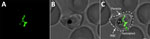
Figure. Visualization of the apicoplast organelle inside a malaria parasite. Microscopy image of a Plasmodium falciparumtransgenic parasite expressing a green fluorescent protein (GFP) fused to a transit peptide, which…
Regardless of photosynthesis, these plastids share similar metabolic pathways, have a small circular remnant genome, and are engulfed by no less than 4 distinct membranes (Figure). Perhaps more than anything, these membranes tell the evolutionary story of the apicoplast; much like a Russian Matryoshka doll, one organism is nested within another.
[ad_2]
Source link