


[ad_1]
Disclaimer: Early release articles are not considered as final versions. Any changes will be reflected in the online version in the month the article is officially released.
Author affiliation: Guangxi Center for Disease Prevention and Control, Nanning, China (J. Wang, F. Bi, X. Luo, H. Huang, W. He, N. Kang, J. Wang, Y. Ju, G. Lan); Guangxi Key Laboratory of Major Infectious Disease Prevention and Control and Biosafety Emergency Response, Guangxi Center for Disease Control and Prevention, Nanning (J. Wang, Y. Ju, G. Lan); Nanning Center for Disease Prevention and Control, Nanning (C. Liang); The First Affiliated Hospital of Guangxi University of Chinese Medicine, Nanning (Y. Zhao)
Since avian influenza virus (AIV) subtype H10 was isolated in 1949, >2,000 H10 subtype AIVs have been isolated from wild waterfowl, poultry, and mammals worldwide (1). Cross-species spillovers make AIV prevention and control a major One Health challenge (2). According to the World Health Organization weekly update on AIV surveillance published December 20, 2024, only 3 human cases of AIV A(H10N3) virus infection had been reported worldwide, all from China (3). We report another human case of H10N3 virus infection in Nanning, Guangxi Zhuang Autonomous Region, China.
On December 12, 2024, a 23-year-old woman began experiencing fever (maximum axillary temperature 40°C) and cough. After failed symptomatic management at a local clinic on December 16, she was referred to the hospital for outpatient evaluations on December 17 and 18. Her condition deteriorated, and she was admitted to the hospital on December 19 with severe community-acquired pneumonia complicated by type I respiratory failure. Moreover, clinical blood and biochemical tests showed elevated C-reactive protein (75.8 mg/L; reference range 0.5–10 mg/L) (Appendix Table).
Chest computed tomography imaging revealed thickened lung markings with patchy areas of high density in both lungs. Because of worsening respiratory failure, the patient was transferred to the respiratory intensive care unit on December 22 for VV-ECMO (venovenous extracorporeal membrane oxygenation) (4).
Figure 1

Figure 1. Timeline of disease progression and treatment history in a case of human infection with avian influenza A(H10N3) virus, China, 2024. BALF, bronchoalveolar lavage fluid; RICU, respiratory intensive care unit; RT-PCR,…
Reverse transcription PCR of sputum specimens analyzed by the Nanning Centers for Disease Control and Prevention were positive for A(H10N3) AIV on December 23. After >10 days of treatment with VV-ECMO and antiviral drugs, the patient recovered uneventfully and was discharged on February 8 (Figure 1).
The patient had no history of exposure to live poultry before disease onset. She worked in a local supermarket’s meat department that processed and sold fresh pork, beef, and poultry products (chicken and duck), but no live poultry was handled on site. Four close contacts and 12 colleagues of the case-patient completed a 10-day health monitoring period and underwent influenza nucleic acid testing, all negative for H10N3 virus.
Comprehensive environmental surveillance was conducted across critical exposure sites. Swab samples were collected from the patient’s residence (n = 8); occupational environment, poultry supply chain facilities, and network nodes along the poultry supply chain (n = 61); farmers markets adjacent to the patient’s residence (n = 40); and epidemiologically linked locations visited 10 days before symptom onset (n = 23). Among the 132 environmental samples collected, 73 (55.3%) were positive for pan–influenza A virus; subtypes H9 (42.5%, 31/73) and H5 (8.2%, 6/73) predominated. H10 subtype was not detected in any of the samples.
We obtained whole-genome virus sequences isolated from bronchoalveolar lavage fluid and passaged on embryonated chicken eggs. We designated the virus A/Guangxi/01591/2024/H10N3 (GX01591) and submitted full-length sequences of the polymerase basic (PB) 2 (2,341 nt length), PB1 (2,341 nt), polymerase acidic (PA) (2,233 nt), hemagglutinin (HA) (1,728 nt), nucleoprotein (1,565 nt), neuraminidase (NA) (1,452 nt), matrix (M) (1,027), and nonstructural (890 nt) genes to GISAID (https://www.gisaid.org; accession nos. EPI4019311–8). The egg passage sequence was 100% identical to the original sequence.
Figure 2
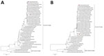
Figure 2. Phylogenetic analysis of avian influenza A(H10N3) virus from human infection, China, 2024. A) Hemagglutinin gene; B) neuraminidase gene. Black triangles indicate A/Guangxi/ 01591/2024(H10N3) virus from patient isolates. Purple circles indicate…
We used EpiFlu BLAST from the GISAID influenza database for sequence alignment and downloaded reference sequences. We performed amino acid site analysis with BioEdit 7.01 software (https://thalljiscience.github.io) and constructed a phylogenetic tree with MEGA version 7 (https://www.megasoftware.net). Phylogenetic analysis revealed that the internal genes of GX01591 were closely related to those of A/Yunnan/0110/2024(H10N3) and belonged to the Eurasian AIV lineage (Figure 2). Evolutionary analysis revealed that the HA genes were reassorted from avian-origin H10Nx, NA genes were reassorted from H7N3, and the other 6 internal genes were reassorted from H9N2 influenza viruses (Appendix Figures 1–6).
The HA proteins contained PEIIQGR↓GLLG at the cleavage site, indicating low pathogenicity. The virus had the QSG motif at the receptor binding site (nucleotide positions 226–8), suggesting avian-like receptor specificity (5). In addition, residues 95Y, 151W, 183H, 190E, 191K, and 194L of the HA protein indicated that the H10N3 virus could bind to avian-like receptors (6). The E119G, H274Y, and R292K molecular markers of NA inhibitors in the NA protein (7), and I38T/M/F of the PA inhibitor in the PA protein exhibited no mutations, suggesting susceptibility to NA inhibitors oseltamivir, zanamivir, and peramivir, and to the PA inhibitor baloxavir. We detected an S to N mutation at residue 31 in the M2 protein, indicating resistance to adamantanes. The molecular markers N30D and T215A in the M1 protein and P42S and V149A in the nonstructural 1 protein exhibited mutations, suggesting increased virulence in mice. We also detected the mammalian adaptive mutation D701N in PB2.
As noted in previously reported human infections caused by H10N3 virus (8,9), our patient initially experienced upper respiratory symptoms before severe pneumonia and respiratory failure developed. Compared with other patients, our patient’s short hospitalization could be attributable to younger age and absence of chronic diseases. GX01591, like other human H10N3 viruses, is an avian-origin reassortant variant (10) and has characteristic avian-like receptor specificity, consequently exhibiting low zoonotic transmission risk. However, the AIV H10 virus subtype should be monitored for potential zoonotic or reassortant viruses.
Mrs. Wang is an epidemiologist at Guangxi Center for Disease Prevention and Control, Nanning, China. Her primary research interest is the prevention and control of acute respiratory infectious disease. Mr. Bi is a microbiologist at Guangxi Center for Disease Prevention and Control, Nanning, China. His primary research interest is the laboratory detection of viral pathogens.
[ad_2]
Source link