



Disclaimer: Early release articles are not considered as final versions. Any changes will be reflected in the online version in the month the article is officially released.

Author affiliation: Friedrich-Loeffler-Institut, Federal Research Institute for Animal Health, Greifswald-Insel Riems, Germany (C. Mehl, J. Sehl-Ewert, R.G. Ulrich); German Centre for Infection Research, Partner Site Hamburg-Lübeck-Borstel-Riems, Greifswald-Insel Riems (C. Mehl, R.G. Ulrich); German Federal Institute for Risk Assessment, Berlin, Germany (K. Schilling-Loeffler, R. Johne); Freie Universität Berlin, School of Veterinary Medicine, Institute of Veterinary Pathology, Berlin (A.F.H. Haake, L. Mundhenk); Microbiological Diagnostics, German Cancer Research Center, Heidelberg, Germany (K. Schmidt); National Institute for Public Health and the Environment, Bilthoven, the Netherlands (M.P. de Cock, M. Maas); Public Health Agency of Lower Saxony, Hannover, Germany (J. Dreesman); University of Bern, Institute of Ecology and Evolution, Bern, Switzerland (G. Heckel); Institute of Biotechnology, Life Sciences Center, Vilnius University, Vilnius, Lithuania (R. Petraityte-Burneikiene)
Despite being closely associated with human settlements for much of recent human history, brown rats (Rattus norvegicus) were not domesticated as pets until the 17th and 18th Centuries in Japan (1). Wild brown rats, which are prolific synanthropic pests, harbor a great diversity of zoonotic agents (2,3). Pet rats, the domesticated form of brown rats, have likewise been associated with outbreaks of Leptospira spp. (4), Seoul virus (SEOV; Orthohantavirus seoulense) (5), and cowpox virus (6).
SEOV is a zoonotic hantavirus found in both wild and pet rats globally (7). In Germany, pet rats have been linked to 4 detections of cases of human SEOV infection (5,8,9). Additional zoonotic pathogens in wild rats include the human pathogenic hepatitis E virus (HEV; Paslahepevirus balayani) and rat hepatitis E virus (rHEV; Rocahepevirus ratti). HEV is one of the main causative agents of acute hepatitis in humans worldwide and is typically transmitted through the fecal–oral route, consumption of undercooked pork, or blood and organ transplants. rHEV has been detected in wild brown rats and black rats (Rattus rattus) worldwide and is now recognized as a novel human pathogen causing acute and chronic hepatitis (10,11). Rats (Rattus spp.) are reservoirs of rHEV but have also been reported to be incidentally infected with genotype HEV-3 (12).
To assess the presence of rHEV, HEV-3, and other related hepeviruses, as well as potential co-infection with SEOV and tissue distribution of hepevirus RNA and SEOV antigen in SEOV-infected and noninfected pet rats, we screened samples from 13 pet rats associated with 3 SEOV detections in human patients in Germany by molecular and serologic assays. Of those 13 pet rats, 11 tested positive for SEOV RNA in previous studies (5,9) (Appendix Table 1). We extracted RNA from liver tissue and screened it by a nested broad-spectrum reverse transcription PCR (RT-PCR) capable of detecting multiple HEV genotypes and rHEV (Appendix). By sequencing the RT-PCR products, we identified rHEV RNA in 3 of the 13 pet rats (KS19/1354, KS19/2196, and KS22/001), a finding further confirmed by rHEV-specific real-time RT-PCR (Appendix Table 1). The 3 pet rats, which were previously shown to be infected by SEOV (Appendix Table 1), came from 3 different owners; 2 rats were obtained from the same vendor and the source for the third rat was unknown (5,8,9). The 276 nt-long open reading frame (ORF) 1 and corresponding amino acid (aa) sequences of rHEV from the 3 pet rats were nearly identical (99.15%–100%) (Appendix Table 2).
Figure
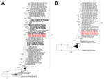
Figure. Phylogenetic relationship of rat hepatitis E virus (rHEV) sequences determined in study of pet rats from Germany co-infected with Seoul hantavirus and wild brown rats from the Netherlands with published…
For further sequence comparisons and phylogenetic analyses, we downloaded 91 partial and 60 complete genome rHEV sequences from GenBank (Figure). In addition, we determined short ORF1 sequences of 12 wild brown rats from Rotterdam (n = 8) and Amsterdam (n = 4) (the Netherlands) that previously tested positive for rHEV RNA (2). Phylogenetic analysis demonstrated that the 3 pet rat–derived sequences were highly similar (98.55%–98.91%) to a wild rat–derived rHEV sequence from Rotterdam (M226) (Figure, panel A).
A multiplex serology approach using bead-based Luminex technology (R&D Systems, https://www.rndsystems.com) (Appendix) detected rHEV-reactive antibodies in 12 of 13 pet rat blood samples; 3 of those 12 samples also showed reactivity to HEV-3 antigen (Appendix Table 1). Subsequent Western blot analysis using the same 2 antigens mainly supported multiplex serology results: 11 of 12 multiplex serology–positive samples also tested positive for rHEV. Of the 11 seropositive samples, 10 also reacted in the Western blot assay with the HEV-3 antigen (Appendix Table 1, Figure 1). That high seroprevalence suggests widespread circulation in pet rat breeding facilities, which should be confirmed using a larger sample size. In all 3 rHEV RNA–positive pet rats, rHEV-reactive antibodies were detected by multiplex serology. Differences in the results of multiplex serology and Western blot assay can be explained by differences in the epitope presentation and sensitivity of both assays.
We obtained near-full rHEV genome sequences from the 3 pet rats from Germany (KS19/1354, KS19/2196, and KS22/001) and from 3 of the wild brown rats from the Netherlands by using a combination of high-throughput sequencing and RT-PCR–based sequence determination of the hypervariable genome region. The wild brown rats (M226, M268, and M186) from Rotterdam were selected for different levels of ORF1 sequence similarity to the 3 pet rat–derived sequences (Figure, panel A). The rHEV full genome sequences from the 3 pet rats were >99.7% identical, and nucleotide and amino acid sequences of all 4 ORFs and encoded proteins were >99.2% identical (Appendix Table 2). As in ORF1, the full genomes from the 3 German pet rats were most similar to the sequence from wild brown rat M226 from Rotterdam (full sequence >99.2% identical; each ORF >98.3%) (Appendix Table 3) and clustered with that sequence in the phylogeny (Figure, panel B). The most closely related rHEV sequences from Germany were detected in wild brown rats from Hamburg (Figure, panel B). The only sequence from a human patient in Germany (13) clustered among sequences from wild brown rats from Berlin, separate from the pet rat sequences (Figure, panel B; Appendix Table 3).
On the basis of the complete coding sequences of pet rat–derived rHEV, we selected probe sequences to detect rHEV RNA through in situ hybridization in formalin-fixed, paraffin-embedded tissues of the pet rats mounted on adhesive microscope slides (Appendix). We clearly detected a disseminated in situ hybridization signal using brightfield microscopy in the livers (hepatocytes) of rats KS19/1354, KS19/2196, and KS22/001, confirming the rHEV quantitative RT-PCR results (Appendix Table 1, Figure 2). In addition, we detected an oligofocal in situ hybridization signal in the spleen of pet rat KS22/001 and in the intestinal lumen of pet rat KS19/1354. We detected no signal in the other tissues tested (Appendix Table 4).
Nucleocapsid protein immunohistochemistry analysis of the rHEV-SEOV coinfected pet rats KS19/1354, KS19/2196, and KS22/001 demonstrated SEOV antigen in multiple organs, predominantly the kidney and brain; antigen was detected less frequently in the spleen, intestine, liver, and lung (Appendix Tables 1, 3, Figure 2). That distribution is consistent with the previously described widespread endothelial tropism of SEOV in naturally infected wild rats and experimentally infected laboratory rats (14). In our study, advanced autolysis limited the assessment of pathological changes. We could not assess colocalization of both viruses because of insufficient tissue material.
The co-infection of pet rats with the zoonotic pathogens SEOV and ratHEV and the rat-to-human transmission of SEOV in those cases (8,9) highlights a public health concern posed by pet rats. Unfortunately, the owners of the pet rats did not consent to providing blood samples; therefore, their rHEV serologic status could not be assessed.
The near-identical rHEV sequences in the pet rats indicate a single incursion event consistent with potentially being independently obtained from the same vendor in Germany (5). The close similarity between the pet rat rHEV sequences and a wild rat–derived sequence from the Netherlands might indicate a wild rat origin of pet rat rHEV, further circulation by pet rat trade, and (at least mostly) uncoupled dynamics of rHEV in wild and captive rats. Owners and general practitioners should be aware of potential zoonotic risks associated with pet rats so that preventive hygienic measures can be taken and correct diagnosis supported.
Dr. Mehl is a postdoctoral researcher at the Friedrich-Loeffler-Institut in Greifswald-Insel Riems, Germany. His research interests include the interplay between pathogen and host ecology, with a focus on small mammals.
We thank Dörte Kaufmann, Stephan Drewes, Elisabeth Striese, Stephanie Hübl, Silvia Schuparis, Christiane Klier, and Simon Dökel for their support.
Ethical approval was not required for this investigation because it was conducted under the German Protection against Infection Act with the aim of investigating transmission routes and halting further spread of infection.
All novel sequences were uploaded to GenBank (accession nos. PZ581217–22 and PZ583560–71).
Funding was provided through the German Center for Infection Research, Thematical Translation Unit “Emerging Infections” (grant no. 01.808_00). C.M. acknowledges intramural funding by Friedrich-Loeffler-Institut, and R.J. acknowledges intramural funding by German Federal Institute for Risk Assessment.