



Sample Collection and Bat Species Identification
Figure 1

Figure 1. Bat collection sites for study of zoonotic Pteropine orthoreovirus in Rousettus amplexicaudatusfruit bats, Indonesia. Map shows Central Java (Surakarta city and Magelang Regency), the Special Region…
We collected a total of 182 bats during 2 sampling periods (126 in 2020 and 56 in 2021) from animal markets and bat suppliers operating in several locations across Central Java (Surakarta City and Magelang Regency), Yogyakarta Special Region, and West Java (Bogor City and Cianjur Regency) (Figure 1). We taxonomically classified the collected bats into 4 species: Cynopterus brachyotis (n = 45), Macroglossus minimus (n = 5), R. amplexicaudatus (n = 96), and Pteropus vampyrus (n = 36).
Virus Isolation, WGS, and Bioinformatics Analysis
Figure 2
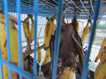
Figure 2. Rousettus amplexicaudatus bats, a frugivorous species collected during study of zoonotic Pteropine orthoreovirus in R. amplexicaudatus fruit bats, Indonesia.
Two virus isolates, designated PRV/SJD-K30/ID/2021 and PRV/SJD-K50/ID/2021, exhibited CPEs in Vero cell cultures. Therefore, we conducted further analyses of those isolates. Both samples were derived from fruit bats identified morphologically as R. amplexicaudatus (Figure 2). Those 2 bats were obtained from a bat collector operating in a residential area in Cianjur, West Java, Indonesia, where bats are sold for human consumption.
We screened samples for influenza A virus, bat coronavirus RdRp and spike genes, and SARS-CoV-2 N1 and N2 gene regions (Table 1). All assays were negative except for the bat coronavirus RdRp screening assay, which yielded positive results in both PRV/SJD-K30/ID/2021 and PRV/SJD-K50/ID/2021. We obtained PRV/SJD-K30/ID/2021 from a live male bat (live at the time of sampling) and PRV/SJD-K50/ID/2021 from a dead male bat; both isolates showed identical screening results (Table 2). Both isolates yielded positive results in the bat coronavirus RdRp screening assay and produced CPE in Vero cell cultures. However, next-generation sequencing did not confirm coronavirus genomic sequences in the samples. Instead, sequence analysis identified a novel PRV, indicating the initial RdRp screening by RT-PCR produced nonspecific amplification that was not corroborated by WGS.
Figure 3

Figure 3. Schematic representation of the complete orthoreovirus virion and 9 genomic segments sequenced in study of zoonotic Pteropine orthoreovirus in R. amplexicaudatusfruit bats, Indonesia. Both PRV/SJD-K30/ID/2021 and…
We reconstructed genomes of the isolated PRVs according to WGS results (Figure 3). PRV genomes comprise 10 double-stranded RNA segments that are classified into 3 large (L), 3 medium (M), and 4 S segments. Each segment encodes >1 protein essential for virus replication, structure, or host interaction. However, in our study, we only recovered 9 segments, indicating incomplete genome acquisition possibly caused by low RNA abundance, degradation, or technical limitations during amplification or sequencing. S1 was not recovered. We further assessed recovery by nested PCR targeting S1 and S2 gene segments by using RNA extracted from Vero cell culture isolates at passage numbers 0 and 1. Although the S2 segment was successfully amplified, the S1 segment yielded only a weak and barely detectable amplicon (Appendix Figure 1). The weak S1 amplification is consistent with low template abundance and might explain the inability to recover all genome segments during next-generation sequencing.
We reconstructed the orthoreovirus virions, which contained 9 genomic segments ranging in size from 1,076 to 3,843 bp (Figure 3). The absence of segment S1, commonly associated with the σC protein involved in cell attachment and host specificity, might have affected downstream phenotypic assessments but did not hinder classification of the isolates within the PRV group. Comparative alignment of segment sizes and gene arrangements further supports the identity of the isolated viruses as a member of the PRV species.
Figure 4

Figure 4. Maximum-likelihood phylogenetic tree of Pteropine orthoreovirus genomic L1 segment found in Rousettus amplexicaudatusfruit bats, Indonesia. Black circles indicate orthoreovirus isolates PRV/SJD-K30/ID/2021 and PRV/SJD-K50/ID/2021 identified in this…
Figure 5

Figure 5. Maximum-likelihood phylogenetic tree of Pteropine orthoreovirus genomic L2 segment found in Rousettus amplexicaudatusfruit bats, Indonesia. Black circles indicate orthoreovirus isolates PRV/SJD-K30/ID/2021 and PRV/SJD-K50/ID/2021 identified in this…
Figure 6

Figure 6. Maximum-likelihood phylogenetic tree of Pteropine orthoreovirus genomic L3 segment found in Rousettus amplexicaudatusfruit bats, Indonesia. Black circles indicate orthoreovirus isolates PRV/SJD-K30/ID/2021 and PRV/SJD-K50/ID/2021 identified in this…
Figure 7

Figure 7. Maximum-likelihood phylogenetic tree of Pteropine orthoreovirus genomic M1 segment found in Rousettus amplexicaudatusfruit bats, Indonesia. Black circles indicate orthoreovirus isolates PRV/SJD-K30/ID/2021 and PRV/SJD-K50/ID/2021 identified in this…
Figure 8

Figure 8. Maximum-likelihood phylogenetic tree of Pteropine orthoreovirus genomic M2 segment found in Rousettus amplexicaudatusfruit bats, Indonesia. Black circles indicate orthoreovirus isolates PRV/SJD-K30/ID/2021 and PRV/SJD-K50/ID/2021 identified in this…
Figure 9

Figure 9. Maximum-likelihood phylogenetic tree of Pteropine orthoreovirus genomic M3 segment found in Rousettus amplexicaudatusfruit bats, Indonesia. Black circles indicate orthoreovirus isolates PRV/SJD-K30/ID/2021 and PRV/SJD-K50/ID/2021 identified in this…
Figure 10

Figure 10. Maximum-likelihood phylogenetic tree of Pteropine orthoreovirus genomic S2 segment found in Rousettus amplexicaudatusfruit bats, Indonesia. Black circles indicate orthoreovirus isolates PRV/SJD-K30/ID/2021 and PRV/SJD-K50/ID/2021 identified in this…
Figure 11

Figure 11. Maximum-likelihood phylogenetic tree of Pteropine orthoreovirus genomic S3 segment found in Rousettus amplexicaudatusfruit bats, Indonesia. Black circles indicate orthoreovirus isolates PRV/SJD-K30/ID/2021 and PRV/SJD-K50/ID/2021 identified in this…
Figure 12

Figure 12. Maximum-likelihood phylogenetic tree of Pteropine orthoreovirus genomic S4 segment found in Rousettus amplexicaudatusfruit bats, Indonesia. Black circles indicate orthoreovirus isolates PRV/SJD-K30/ID/2021 and PRV/SJD-K50/ID/2021 identified in this…
We created phylogenetic trees for each of the 9 gene segments (Figures 4,5,6,7,8,9,10,11,12). Phylogenetic analyses of segments L1–L3, M1–M3, and S2–S4 showed that PRV/SJD-K30/ID/2021 and PRV/SJD-K50/ID/2021 consistently clustered with other bat-associated orthoreoviruses, including Nelson Bay (China), Cangyuan (China), Melaka (Malaysia), Kampar (Malaysia), and Pulau orthoreoviruses, as well as PRV (Malaysia) and PRV Garut-69 (Indonesia). Phylogenetic trees of L2, L3, M1, M3, S2, and S3 segments indicated that PRV/SJD-K30/ID/2021 and PRV/SJD-K50/ID/2021 were most closely related to the PRV Garut-69 strain isolated from Pteropus vampyrus bats, which was previously associated with human infections in Indonesia.
We generated heat maps to compare the 9 segments of PRV/SJD-K30/ID/2021 and PRV/SJD-K50/ID/2021 with sequences from other viruses (Appendix Figures 2–10). L1 segments exhibited 100% nucleotide identity with each other. Both isolates showed the highest similarity to PRVs, particularly Pulau (94.6%) and Melaka (94.1%) reoviruses, followed by Cangyuan orthoreovirus (91.7%). In contrast, identities with avian, reptile, and non-PRV mammal orthoreoviruses were much lower (≈27%–35%). The L1 sequences firmly place the 2 isolates within the PRV group. L2 gene segments, which encode capsid proteins, were 100% identical between the 2 isolates. Both isolates showed the highest identities with Pulau (93.3%) and Melaka (92.5%) reoviruses, followed by Cangyuan orthoreovirus (91.6%). Non-PRV reference sequences showed 65% identity. Thus, the L2 segment demonstrated a highly conserved backbone consistent with the PRV lineage. The L3 gene segments of the 2 isolates were 100% identical. The closest reference sequences were Melaka (78.9%), Pulau (78.7%), and Cangyuan (78.1%) orthoreoviruses, whereas other orthoreoviruses showed only 27%–50% identity. Those findings indicate the L3 segment of the 2 isolates clusters tightly within the PRV group.
M1 gene segments from PRV/SJD-K30/ID/2021 and PRV/SJD-K50/ID/2021 shared 99.1% identity. We detected the highest external similarities between PRV/SJD-K30/ID/2021 and Pulau (77.3%), Melaka (77.1%), and Cangyuan (77.0%) orthoreoviruses. PRV/SJD-K50/ID/2021 showed lower similarities with Pulau (76.8%), Melaka (76.5%), and Cangyuan (76.4%) orthoreoviruses. Those findings suggest the M1 segment has strong PRV affinity and shows minimal divergence between the 2 isolates from Indonesia.
The M2 gene segments, which encode outer capsid proteins, were 100% identical between the 2 isolates. The closest matches were Pulau (82.2%) and Melaka (82.0%) orthoreoviruses, whereas the match with Cangyuan orthoreovirus was slightly lower (69.8%). That pattern suggests indicates the conserved outer capsid determinants of the 2 isolates are similar to those of Pulau and Melaka viruses. In addition, the M3 segment of the 2 isolates shared 100% identity. The highest sequence similarities were with Pulau and Melaka orthoreoviruses (85.8% each), followed by Cangyuan orthoreovirus (85.0%). Thus, the M3 segment is strongly conserved within PRVs.
Unlike the other segments, the nucleotide identity of S2 gene segments between PRV/SJD-K30/ID/2021 and PRV/SJD-K50/ID/2021 decreased to 88.9%. Nevertheless, both isolate sequences remained closest to other PRVs, including Pulau (83.3%), Melaka (81.5%–81.8%), and Cangyuan (81.7%) orthoreoviruses. The decreased identity between the 2 isolates likely indicates an intra-PRV reassortment event in the S2 segment. However, the presence of numerous ambiguous nucleotide positions in 1 of the sequences might have influenced the estimated sequence identity and limited the strength of that inference.
The S3 gene segments of the 2 isolates were nearly identical (99.9%). The highest similarities were to Pulau (78.1%–78.2%), Melaka (77.0%), and Cangyuan (76.3%–76.4%) orthoreoviruses. No evidence of reassortment was observed in this segment.
The S4 gene segments of the 2 isolates were 100% identical. The highest identities were with Melaka orthoreovirus (86.7%), followed by Cangyuan (85.2%) and Pulau (83.3%) orthoreoviruses, suggesting that the S4 segment, which plays a role in viral entry, is highly conserved among PRVs.
Taken together, analyses of all 9 segments consistently place PRV/SJD-K30/ID/2021 and PRV/SJD-K50/ID/2021 within the PRV clade, and the closest affinities were to Melaka, Pulau, and Cangyuan orthoreoviruses. The 2 isolates from Indonesia were nearly identical across the genome (>99%–100%), except for the S2 segment, which declined to ≈89%. That pattern strongly suggests reassortment within locally circulating PRVs that specifically involves the S2 segment, which encodes an inner capsid structural protein. Our findings support the existence of active PRV circulation in Indonesia with >2 divergent S2-associated lineages embedded in an otherwise conserved PRV genomic background.