



Disclaimer: Early release articles are not considered as final versions. Any changes will be reflected in the online version in the month the article is officially released.
Author affiliation: Hokkaido University, Sapporo, Japan (Y. Qiu, P.A. Jiménez, A. Ohnuma, N. Nonaka, R. Nakao); Kitasato University, Towada, Japan (Y. Qiu, T. Kakuda); Tokai University, Sapporo (K. Kawai); The Asian Bat Research Institute, Nara, Japan (H. Tamura); Saitama Medical University, Moroyama, Japan (M. Takahashi); Zoukei Co., Ltd., Obihiro, Japan (S. Kamiyama)
Scrub typhus, or tsutsugamushi disease, is a vectorborne zoonosis caused by Orientia tsutsugamushi bacteria. O. tsutsugamushi is transmitted by larval trombiculid mites, or chiggers, and is primarily maintained within chigger populations through transovarial and transstadial transmission (1). Ecologic studies of scrub typhus have largely focused on rodent-associated Leptotrombidium chiggers, which are the principal vectors in many endemic areas (1). In Japan, where ≈300–500 scrub typhus cases are reported annually (2), the ecologic diversity of O. tsutsugamushi in wildlife-associated chiggers remains poorly understood.
Recent molecular detection of O. tsutsugamushi DNA in bats from the China–Myanmar border raised questions about the roles of bats and their ectoparasites in Orientia ecology (3). Bat-associated chiggers are diverse and widely distributed (4), but their involvement in Orientia ecology remains limited. We report the isolation and propagation of O. tsutsugamushi from Whartonia prima chiggers collected on Yonaguni Island, Japan, from Hipposideros turpis, a cave-dwelling insectivorous bat endemic to the Yaeyama Islands (5).
We collected larval chiggers from H. turpis bats on Yonaguni Island in September 2024 and 2025. Bat capture and handling were approved by the Ministry of the Environment, Tokyo, Japan (permission nos. 2402141 and 2502282), and complied with relevant institutional and national guidelines. We examined a total of 80 H. turpis bats, yielding 212 chiggers: 31 in 2024 and 181 in 2025. We identified all chigger mites morphologically as W. prima by using taxonomic descriptions and keys (6,7). We used the chigger mites collected in 2024 for molecular screening. Of the 181 chigger mites we collected in 2025, we used 37 mites for molecular screening, 129 mites for isolation attempts, and 4 mites for scanning electron microscopy; we excluded 11 mites because of poor specimen condition.
Figure 1
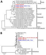
Figure 1. Phylogenetic analyses of Orientia tsutsugamushi detected and isolated from bat-associated Whartonia primachiggers, Yonaguni Island, Japan. A) Maximum-likelihood tree on the basis of full-length 56-kDa type-specific antigen…
We tested 68 chigger mites individually for O. tsutsugamushi by using real-time PCR targeting the 16S rRNA gene (8) and nested PCR targeting the 56-kDa type-specific antigen (TSA56) gene (9). We sequenced amplicons from all nested PCR-positive chigger mite samples, revealing 2 partial TSA56 genotypes. We amplified full-length O. tsutsugamushi TSA56 from 10 mite samples representing both partial genotypes by using previously described primers (Appendix) (10). Of the 68 chigger mites tested, 64 (94%, 95% CI 86%–98%) were positive for O. tsutsugamushi by real-time PCR, and 55 (81%, 95% CI 70%–89%) were positive for O. tsutsugamushi by nested PCR. We obtained full-length TSA56 sequences from 2 of the 10 selected chigger mite samples; both clustered within the Karp-related lineage (Figure 1, panel A).
For isolation, we pooled the 129 W. prima chiggers collected in 2025 by individual bat host. Each pool consisted of 2–5 chiggers from the same bat and was homogenized in sucrose–phosphate–glutamate buffer. We inoculated the homogenates onto L929 cells maintained in Leibovitz’s L-15 medium supplemented with 2% fetal bovine serum without antimicrobials. We passaged the cultures every 2 weeks and monitored O. tsutsugamushi DNA by using real-time PCR at each passage. We maintained uninfected L929 cell controls in parallel that remained negative for O. tsutsugamushi. We discarded contaminated cultures immediately. Of 28 chigger pools, 4 pools yielded O. tsutsugamushi isolates: Donan 18-3R (3 chiggers), Donan 17-2L (2 chiggers), Donan 14-1L (5 chiggers), and Donan 14-2R (5 chiggers). Three cultures remained negative, and the remainder were excluded because of contamination. We maintained all 4 isolates through 4 passages and stored fourth-passage infected cell suspensions in sucrose–phosphate–glutamate buffer.
We generated full-length TSA56 gene sequences for 2 O. tsutsugamushi isolates, Donan 18-3R and Donan 17-2L, and compared them with sequences obtained directly from selected PCR-positive W. prima chiggers (GenBank accession nos. LC896643, LC896644, LC939506, and LC939507) (Figure 1, panel A). Partial TSA56 sequences from the remaining O. tsutsugamushi isolates, Donan 14-1L and Donan 14-2R (accession nos. LC939530 and LC939531), were identical to the corresponding regions of the full-length sequences from Donan 18-3R and Donan 17-2L. We performed multilocus sequence analysis (MLSA) on the basis of 11 housekeeping genes for 10 chigger samples selected for full-length TSA56 analysis and 2 O. tsutsugamushi isolates, Donan 18-3R and Donan 17-2L, by using primers described previously (11) (accession nos. LC897744–55 and LC939508–29). The concatenated MLSA sequences of Donan 18-3R and Donan 17-2L were identical. The O. tsutsugamushi isolate sequences and sequences obtained directly from PCR-positive chigger samples clustered within the O. tsutsugamushi clade in MLSA, supporting their identification as O. tsutsugamushi (Figure 1, panel B).
Figure 2
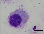
Figure 2. Microscopy of Orientia tsutsugamushi isolate Donan 18-3R detected and isolated from bat-associated Whartonia primachiggers, Yonaguni Island, Japan. Giemsa-stained L929 cells 5 days after inoculation with fourth-passage…
Figure 3
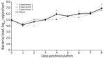
Figure 3. Growth kinetics of Orientia tsutsugamushi isolate Donan 18-3R detected and isolated from bat-associated Whartonia primachiggers, Yonaguni Island, Japan. Growth kinetics were assessed by quantitative PCR and…
To characterize a representative isolate, we inoculated fourth-passage Donan 18-3R onto fresh L929 cells. We identified numerous punctate, basophilic structures in the cytoplasm outside the nucleus from Giemsa-stained cells 5 days postinoculation (dpi), a pattern consistent with intracellular O. tsutsugamushi (Figure 2). For growth analysis, we inoculated fourth-passage Donan 18-3R onto L929 cells in 24-well plates. We extracted DNA daily from 1 well and calculated bacterial copy numbers by using quantitative PCR, which we independently performed 3 times. After an initial decrease at 1 dpi, mean +SD bacterial loads increased from 4.19 +0.21 log10 copies/well at 1 dpi to 6.93 +0.59 log10 copies/well at 8 dpi, demonstrating growth of Donan 18-3R in L929 cells (Figure 3).
We provided evidence that bat-associated W. prima chiggers can harbor viable O. tsutsugamushi, demonstrated by isolation, serial propagation, microscopic examination, and growth kinetics. The O. tsutsugamushi isolates clustered phylogenetically within the Karp-related lineage.
Although vector competence, vertebrate host involvement, and human exposure were not demonstrated, isolation of viable O. tsutsugamushi from bat-associated chiggers demonstrates bacterial viability, which cannot be inferred from molecular detection alone. In the Miyako Islands of the southern Ryukyu Archipelago, Japan, Leptotrombidium deliense mites are implicated in the transmission of O. tsutsugamushi causing human scrub typhus (12). A Karp-related strain was reported from a human scrub typhus case on Ikema Island in the Miyako Islands, Japan, although it was not predominant among genotyped cases (12). In contrast, O. tsutsugamushi was not reported previously in the literature from animals on Yonaguni Island or elsewhere in the Yaeyama Islands, Japan, and no human scrub typhus cases have been reported from that region to our knowledge. We identified a previously unrecognized ecologic setting for O. tsutsugamushi in the southern Ryukyu Archipelago, Japan, suggesting more diverse regional maintenance.
W. prima chiggers have been reported from several bat species across East and Southeast Asia (4,13). However, W. prima chigger strict host specificity has not been established, and knowledge of this species’ role in O. tsutsugamushi transmission to vertebrates remains limited. The broad geographic distribution of W. prima chiggers suggests similar bat-associated chigger systems might occur elsewhere. Together with recent detection of O. tsutsugamushi DNA in bats from the China–Myanmar border (3), our findings raise the possibility that bat–ectoparasite interfaces represent an underrecognized component of Orientia ecology. Additional studies are needed to clarify bacterial localization in chiggers, vector competence, vertebrate host involvement, human exposure, and public health relevance. Because the W. prima chigger larvae examined in this study were collected from bats, detection and preferably isolation of O. tsutsugamushi from unfed W. prima larvae will be needed to confirm transovarial transmission. Our findings expand the known ecologic settings of viable O. tsutsugamushi and highlight bat–chigger interfaces as an underrecognized component of Orientia ecology.
Dr. Qiu is an associate professor at Kitasato University, Towada, Japan. His research focuses on the diversity and maintenance mechanisms of vector-borne bacterial pathogens, including Borrelia, Orientia, and Rickettsia, in natural ecosystems.