



Disclaimer: Early release articles are not considered as final versions. Any changes will be reflected in the online version in the month the article is officially released.
Author affiliation: Friedrich-Loeffler-Institut, Greifswald-Insel Riems, Germany (K. Wernike, B. Hoffmann, M. Eshak, F. Pfaff, D. Höper, M. Beer); State Institute for Chemical and Veterinary Analysis Freiburg, Freiburg, Germany (E.K. Link); Animal Disease Fund Baden-Württemberg, Cattle Health Service, Freiburg (S. Rotheneder)
Figure 1
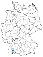
Figure 1. Location of cattle herds within Germany from which blood samples from affected cattle were subjected to metagenomic analysis and which tested positive for the novel Shamonda virus clade (blue dots)…
Since mid-June 2026, farmers and veterinarians in southern Germany and Switzerland have reported an acute disease in dairy cattle characterized by a marked decrease in milk production, diarrhea, fever, reduced activity, and occasionally mild respiratory signs for a short period. By the end of July, >100 affected farms had been reported to the local cattle health service in the German federal state of Baden-Württemberg. At the herd level, the within-herd illness rate was very high (up to 100%), but no fatalities were reported. Given the high environmental temperatures at the time, heat stress was initially considered to be at least a contributing factor. However, an infectious disease was suspected, and a vectorborne virus was thought likely because of the time of year and the rate at which the disease was spreading. Classical endemic and emerging viruses such as bluetongue virus, epizootic hemorrhagic disease virus, Schmallenberg virus (SBV), pestiviruses, bovine herpesvirus type 1, bovine ephemeral fever virus, West Nile virus, and Usutu virus were excluded as the causative agent by real-time reverse transcription PCR (rRT-PCR). To identify the cause of the outbreak, serum and whole blood samples from 4 herds in Baden-Württemberg (Figure 1) with acutely diseased cattle were sent by the local state laboratory to the Friedrich-Loeffler-Institut, Germany’s federal research institute for animal health, for further investigations using generic PCR systems and metagenomic analysis.
In total, a serum and a whole blood sample from 27 individual animals from 4 different cattle herds were submitted. Because of the small volume available per sample, we pooled up to 6 samples according to sample matrix and herd, resulting in 16 pools (2 whole blood pools and 2 serum pools for each of the 4 herds). We inoculated the samples onto monkey kidney (Vero) and baby hamster kidney (BHK-21-BSR/5) cells and isolated infectious virus from sample pools from 3 herds on Vero cells and from 1 of those pools additionally on BHK-21-BSR/5. We also extracted nucleic acid from the sample pools with the King Fisher 96 Flex purification system and the NucleoMag Vet Kit (Macherey Nagel, https://www.mn-net.com) according to the manufacturer’s instructions. Subsequent testing by rRT-PCR for the ruminant orbiviruses bluetongue virus and epizootic hemorrhagic disease virus, morbilliviruses (generic RT-PCR), influenza A and D viruses, bovine ephemeral fever virus, and Hefer Valley virus gave consistently negative results. However, 10 of the 16 pools tested positive by a generic rRT-PCR for viruses of the Simbu serogroup (1), genus Orthobunyavirus (quantification cycle values 19.8–37.6). From every investigated cattle farm, >1 sample pool gave a positive result. We obtained positive results from serum and whole blood. In addition, all but 2 pools tested positive by an rRT-PCR targeting the small segment of the Simbu serogroup virus Sathuperi virus (2) (quantification cycle values 20.5–29.2). In contrast, all those samples tested negative by a specific real-time RT-PCR for SBV (3), the only Simbu serogroup virus known to circulate in Central Europe to date (4,5). Twenty-three of the serum samples tested positive by an SBV antibody ELISA (ID Screen Schmallenberg Virus Competition Multi-species; Innovative Diagnostics, https://www.innovative-diagnostics.com), 1 sample was questionable, and 3 were negative.
For metagenomic sequencing, we extracted RNA from TRIzol supernatants from serum and blood pools by using the RNAdvance Tissue Kit (Beckman Coulter, https://www.beckmancoulter.com) on a KingFisher Duo Prime platform (Thermo Fisher Scientific, https://www.thermofisher.com). For sequencing, we further processed the RNA as described (M. Eschbaumer et al., unpub. data, https://doi.org/10.64898/2026.03.30.713672). We loaded pooled libraries onto Ion 530 chips by using an Ion Chef instrument (Thermo Fisher Scientific) and sequenced them on an Ion S5 XL platform in 400-bp mode. We assembled the obtained datasets with Newbler 3.0 (Roche, https://www.roche.com). We submitted the resulting complete genome sequence to the International Nucleotide Sequence Database Collaboration (http://www.insdc.org; project no. PRJEB123174).
Figure 2
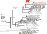
Figure 2. Phylogenetic analysis of the large segment of the novel Shamonda virus clade detected in Germany, 2026. Phylogenetic tree inferred from the amino acid sequence of the RNA-dependent RNA polymerase encoded…
Figure 3
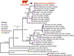
Figure 3. Phylogenetic analysis of the medium segment of the novel Shamonda virus clade detected in Germany, 2026. Phylogenetic tree inferred from the amino acid sequence of the glycoprotein precursor encoded by…
Figure 4
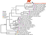
Figure 4. Phylogenetic analysis of the small segment of the novel Shamonda virus clade detected in Germany, 2026. Phylogenetic tree inferred from the amino acid sequence of the nucleocapsid protein encoded by…
The obtained genome showed the highest identity to the Nigeria Shamonda virus (SHAV) isolate Ib An 5550 (GenBank accession no. HE795105.1): 93% nucleotide identity for the large and medium segments and 97% for the small segment. SHAV, which had not been previously detected in Europe, falls within the Simbu serogroup of orthobunyaviruses. We performed phylogenetic analyses separately for each segment. We compared amino acid sequences of the proteins encoded by each segment with 29 reference sequences obtained from the National Center for Biotechnology Information RefSeq database (https://www.ncbi.nlm.nih.gov/refseq) and aligned them by using MUSCLE 5.1 (https://www.drive5.com/muscle5). We inferred maximum-likelihood trees with IQ-TREE 3.1.1 (https://iqtree.github.io) by using the best-fit substitution model. We assessed branch support with 10,000 ultrafast bootstrap and 10,000 Shimodaira–Hasegawa-like approximate likelihood ratio test replicates. We used sequences from the California and Bunyamwera serogroups of orthobunyaviruses as outgroups. All 3 genome segments were most closely related to SHAV of the Simbu serogroup, providing no evidence for an interlineage reassortant origin (Figures 2–4). In addition, the small and large segments were closely related to SBV (Figure 2, 4), whereas the medium segment differed markedly (Figure 3).
The emergence of a second Simbu serogroup virus in the cattle population where SBV has been endemic for many years (6) may have important epidemiologic and veterinary implications. Despite a high seroprevalence against SBV in the affected region (6), the large number of clinical cases associated with the newly detected SHAV clade strongly suggests that existing SBV immunity does not provide meaningful cross-protection. This observation is further supported by the sequence differences in the medium segment (Figure 4), which encodes the viral glycoproteins Gc and Gn, the major antigens of orthobunyaviruses (7). Consequently, population immunity against SBV is unlikely to limit the spread of SHAV in the following summer and autumn months, assuming that SHAV is transmitted by biting insects, as are related Simbu serogroup viruses (4). Moreover, based on observations from other regions where >2 of the vector-transmitted simbuviruses are present (e.g., Australia and the Middle East), the 2 viruses plausibly will establish an alternating or cyclical pattern of circulation, rather than 1 permanently displacing the other (8,9). Such dynamics may be influenced by fluctuations in vector abundance, climatic conditions, and, above all, the level of herd immunity against 1 virus or the other. Long-term surveillance will be essential to determine whether similar epidemiologic patterns will evolve in Europe and to identify factors driving the relative predominance of each virus over time.
In the current outbreak in Europe, acute clinical disease has been reported in cattle in Germany and Switzerland (10). However, the host range of the newly emerged SHAV clade in Europe remains incompletely understood. Although SHAV infections have mainly been documented in Africa and Asia in cattle so far (11,12), infection of other domestic and wild ruminant species cannot be excluded, given the broader range of ruminant hosts observed for several other members of the Simbu serogroup, including SBV (6). Therefore, monitoring sheep, goats, and free-ranging ruminants during the coming months will be crucial to assess the host range. Monitoring animals that were pregnant at the time of infection is particularly important because multiple viruses of the Simbu serogroup, including SHAV and SBV, can cause severe fetal malformations when naive dams are infected during early gestation (3,4,9,12). Such investigations improve understanding of the pathogenesis and ecology of the virus and aid in risk assessments. Continued molecular and serologic surveillance across susceptible species, combined with vector monitoring and identifying the local vector species responsible for virus transmission, will be critical for characterizing the long-term epidemiology of the emerging SHAV clade and for guiding appropriate disease-control measures.
This article was preprinted at https://doi.org/10.64898/2026.08.06.743243.
Drs. Wernike and Hoffmann are veterinarians and senior scientists at the Friedrich-Loeffler-Institut, Greifswald-Insel Riems, Germany. Their research interests focus on emerging viruses, host–virus interaction, diagnostics, and immunoprophylaxis.
We thank Tobias Töws, Patrick Zitzow, Karin Pinger, and Bianka Hillmann for excellent technical assistance.
The samples were taken by veterinarians in the context of health monitoring and as part of their clinical practice; no permissions were needed to collect these specimens.
This study was supported by intramural funding from the Friedrich-Loeffler-Institut, provided by the German Federal Ministry of Agriculture, Food and Regional Identity. This work was further funded by the DURABLE project, co-funded by the European Union, under the EU4Health Programme (project no. 101102733).
The authors declare that the research was conducted in the absence of any commercial or financial relationships that could be construed as a potential conflict of interest.