



Disclaimer: Early release articles are not considered as final versions. Any changes will be reflected in the online version in the month the article is officially released.
Author affiliation: Stanford University, Stanford, California, USA (I.M. Espinal, G.W. Charville, R.A. Brown, K.E. Rieger, S. Fernandez-Pol); Guardant Health, Palo Alto, California, USA (L. Lawrence); Stanford Medicine, Stanford (R.A. Brown, K.E. Rieger)
Trichodysplasia spinulosa polyomavirus (TSPyV) typically causes folliculocentric cutaneous eruptions, usually on the face, although the virus has been detected in tonsillar tissue, blood, urine, cerebrospinal fluid, and respiratory specimens (1–4). Recently, TSPyV was identified within the endothelial cells of tumors from 3 separate patients by analyzing off-target next-generation sequencing reads (5). Subsequently, in situ hybridization demonstrated the localization of TSPyV to intratumoral endothelial cells. The significance of TSPyV within endothelial cells in those cases is uncertain. Of note, all 3 patients received corticosteroids immediately before tumor resection.
We hypothesized that TSPyV might be found in vasculitides or other cutaneous inflammatory conditions. We evaluated skin biopsy samples from 25 patients with inflammatory dermatologic conditions. Of the 25 patients, 13 received corticosteroids before skin biopsy. Among those patients, 10 were treated with corticosteroids within 10 days of the biopsy. Three patients had longer intervals between the corticosteroid dose and the biopsy, with intervals of 108 days, 1,494 days, and 1949 days between the corticosteroid administration and the biopsy. The biopsy samples included leukocytoclastic vasculitis (n = 5), medium-vessel vasculitis (n = 1), interface dermatitis (n = 6), perniosis (n = 2), tumid lupus (n = 1), erythema nodosum (n = 4), calciphylaxis (n = 1), and nonspecific epidermal dermatoses (n =5). In addition, 1 sample of trichodysplasia spinulosa was included as a positive control.
Figure
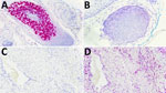
We performed in situ hybridization by using a custom RNAScope probe (5) targeting the complete TSPyV genome on tumor-associated endothelial cells within 25 dermatologic samples representing a range of inflammatory conditions. We used a peptidylprolyl isomerase B RNA probe as a positive control for RNA integrity in all specimens (Figure). We did not detect TSPyV in endothelial cells in any of the 25 inflammatory skin biopsy samples. In contrast, the trichodysplasia spinulosa control had positive staining, and peptidylprolyl isomerase B staining was present in all samples (Table).
We found no evidence of TSPyV infection in endothelial cells in this survey of cutaneous inflammatory conditions, suggesting that TSPyV does not play a role in the cutaneous inflammatory conditions we included. Of note, our cohort is small compared with that of the previous study, which initially led to the identification of TSPyV in 3 cases from 252 specimens (3). Therefore, the prevalence of TSPyV infection of endothelial cells might be too low to be detected in a cohort of only 25 cases. Our findings provide evidence that endothelial TSPyV infection is not commonly detected in inflammatory skin biopsy specimens and helps refine the spectrum of tissues and clinical settings in which TSPyV should be considered.
Dr. Espinal is a fourth-year anatomic and clinical pathology resident at Stanford University and an incoming gastrointestinal pathology fellow. His research interests include gastrointestinal, hematologic, and microbiologic pathology.